

Hologic continuously looks to improve women’s health and enhance patients’ clinical experience. As such, it offers I-View software for Contrast Enhanced 2D (CE2D) imaging on Hologic mammography systems. It can also integrate with the firm’s other technologies, including Genius™ 3D MAMMOGRAPHY™.
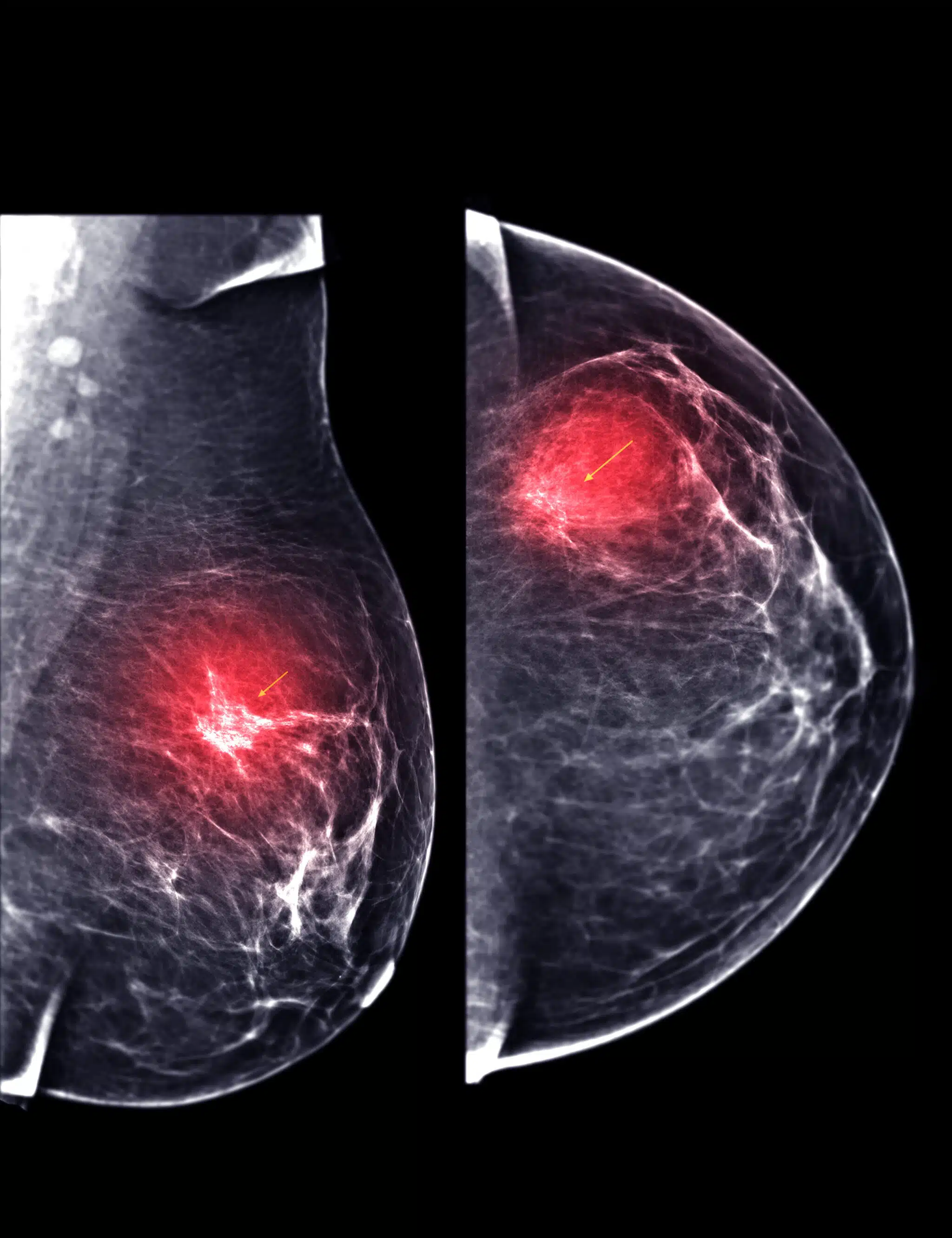
I-View software is a solution that allows enhanced CE2D on Hologic’s mammography systems. Regular CE2D is a technique that uses a contrast agent to highlight areas of abnormal blood flow in the breast, which may indicate cancer.
Agents are a drug type that tends to accumulate where cancer is growing. Successful imaging can enhance diagnostic information to complement 2D and 3D mammography images. It is helpful for women with dense breast tissues or suspicious lesions that are challenging to confirm as cancerous via alternative methods.
Hologic’s I-View software augments this technique by allowing the acquisition of high- and low-energy images in rapid succession and subtracting the background breast tissue to display the contrast-enhanced regions. The solution can optimize image quality and x-ray dose depending on the contrast agents and compression paddles used. Ultimately, the solution gives clinical practitioners more confidence when making diagnoses.
Overview Of Hologic I-View Software Components
Hologic’s I-View software is a sophisticated digital solution for enhancing breast cancer screening and detection. As such, it offers several components to aid clinicians and patients, reducing stress and improving productivity.
Image Acquisition Module
The Image Acquisition Module (IAM) supports 2D and 3D imaging modes, contrast-enhanced spectral mammography (CESM), and synthetic 2D imaging. It controls communication between the mammography system and workstation and images from the detectors and represents the first step in the chain.
Image Processing Module
The Image Process Module (IPM) is where most AI assistance occurs. The unit applies various algorithms to enhance the image quality and optimize how images appear on the screen. Techniques can include:
- Noise sharpening
- Contrast enhancement
- Edge sharpening
- Tissue equalization
- Dual-energy subtraction for CESM images
Image Review Module
The Image Review Module (IRM) lets clinicians review and manipulate outputs on their workstations. It offers tools for image navigation, panning, zooming, annotating, and comparing. The software also allows radiologists to overlay one image on another from third-party modalities, such as MRI or ultrasound, for a complete diagnostic vantage point.
Image Management Module
The Image Management Module (IMM) is critical for image storage and retrieval. It supports various formats for image compression and transmission and can integrate with other systems. It’s also helpful for encryption and upholding patient confidentiality.
Audit trails are also available via this module. Clinicians can track when data entered the system, who accessed it, and when they sent it.
The Benefits Of I-View Software
Hologic’s I-View software offers radiologists and radiography units in hospitals and clinics multiple advantages when diagnosing breast cancer patients. These benefits extend to patients, clinicians, and entire practices.
Faster Workflow And Reporting
First, Hologic’s I-View software streamlines the workflow and reporting process for radiologists, by providing a single workstation for viewing and analyzing 2D and 3D images. It’s also compatible with SecurView DX diagnostic workstation and Affirm Prone biopsy system, which makes it easier to transition from diagnostics to interventions.
I-View also enhances reporting. That’s because it supports the Breast Imaging Reporting and Data System (BI-RADS) classification from the American College of Radiology, helping to standardize results and make clinical findings more comparable. Clinicians can easily share outputs with referring physicians and patients.
Enhanced Lesion Detection
Another benefit of I-View software is the enhanced lesion detection it offers. The tool boosts the contrast and visibility of lesions in the breast tissue across a range of agents, making it easier for radiologists to identify and characterize suspicious findings.
There are also quality-of-life benefits. For instance, I-View software reduces the number of false positives, meaning fewer recalls and invasive biopsies.
Reduced Radiation Exposure
Lastly, Hologic I-View software offers patients reduced radiation exposure. Using synthetic 2D images instead of conventional 2D mammograms cuts the radiation dose significantly without affecting image quality or diagnostic accuracy.
For patients with dense breasts, this feature is helpful. It reduces the need for additional imaging tests and higher radiation doses that might result if they rely on standard 2D mammography.
Conclusion
Hologic I-View software is a robust solution for oncology and breast cancer clinics. It enables contrast-enhanced digital mammography (CEDM) on Hologic 3Dimensions and Selenia Dimensions systems, using iodinated contrast agents to highlight areas of increased blood flow in the breast, which may indicate the presence of cancer.
To learn more about I-View compatible systems, visit Mammo.com, give us a call, or fill out the inquiry request below. We offer many Hologic Mammography systems that have this feature enabled.


